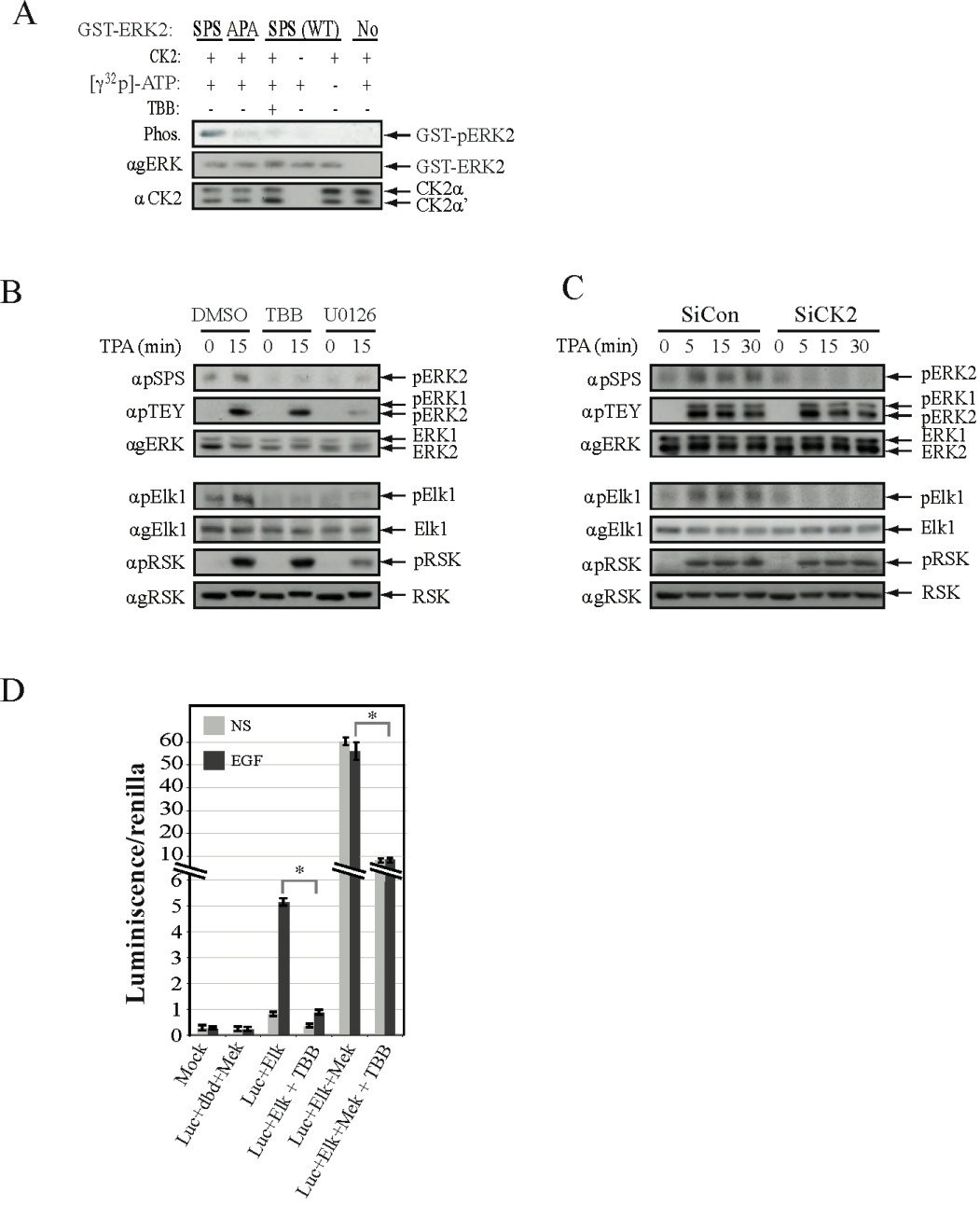
Fig. 2. CK2 phosphorylates ERK's NTS and is involved in the activation of nuclear ERK targets. (A) CK2 phosphorylates ERK's NTS in-vitro. The indicated GST-ERK2 constructs (0.1 mg per sample) were subjected to in-vitro CK2 phosphorylation, that was detected by autoradiography (upper panels). The loaded ERK and CK2 constructs were detected using agERK and aCK2a Abs (middle and lower panels respectively). (B) CK2 inhibition prevents ERK's NTS and Elk1 but not RSK1 phosphorylation. HeLa cells were serum starved (0.1 %, 16 h), pretreated with TBB (10 mM, 2 h), UO126 (5mM; 2 h) or DMSO control, and then stimulated with TPA (250 nM, 15 min) or left untreated as control. Cell extracts were subjected for western blot analysis with the indicated Abs. (C) CK2 knockdown prevents ERK's NTS and Elk1 but not RSK1 phosphorylation. CHO cells were transfected with either siCK2 or non-relevant siRNA (siCon). Thirty six h after transfection, the cells were serum-starved and then either stimulated with TPA (250 nM, 15 min) or left untreated as a control. Cell extracts were subjected to Western blot analysis using the same Abs as in Fig. 2B. Efficiency of CK2 knockdown is shown in Fig.1B. (D) Inhibition of CK2 attenuates the ERK1/2-dependent activation of Elk1. HEK293T cells were transfected with the plasmids: pFC2-Elk1, constitutively active MEK1 (pFC-MEK1), and pFC2-dbd as control. The cells were then either pretreated with TBB (10µM, 2 h), or left untreated as control followed by stimulated with EGF (50 ng/ml) for 6 h where indicated. The graph represents the ratio of the luminescence of pFR-Luc (reporter)/renilla luminescence. The bar-graph is the mean and standard error of three repeats. * P<0.01.